PET CT vs CT Scan: Unveiling the Difference in Medical Imaging
18
0
Guide or Summary:PET and CT scans are two of the most advanced imaging techniques used in medical diagnostics. While both offer invaluable insights into the……
Guide or Summary:

- PET and CT scans are two of the most advanced imaging techniques used in medical diagnostics. While both offer invaluable insights into the human body, they differ significantly in their methodology, applications, and the information they provide to healthcare professionals. Understanding these differences is crucial for patients and healthcare providers alike, as it helps in making informed decisions about which imaging test is most suitable for a particular condition.
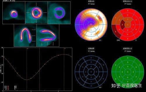
- PET Scan stands for Positron Emission Tomography. This imaging technique involves the injection of a small amount of a radioactive substance, known as a tracer, into the patient's body. As the tracer moves through the body, it is absorbed by metabolically active tissues, which can then be visualized by a PET scanner. The tracer emits positrons, which interact with electrons in the body, producing gamma rays that are detected by the PET scanner. This process allows for the visualization of metabolic activity within the body, making PET scans particularly useful for diagnosing conditions that affect metabolic processes, such as cancer, Alzheimer's disease, and heart disease.
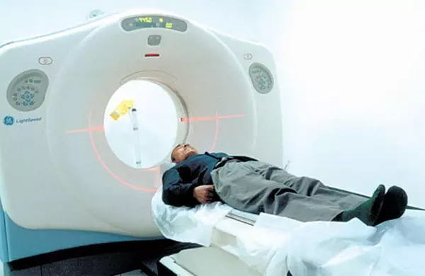
- CT Scan, on the other hand, stands for Computed Tomography. This imaging technique uses X-rays and computer technology to create detailed images of the body's internal structures. During a CT scan, the patient is positioned inside a doughnut-shaped scanner, which rotates around the body, taking multiple X-ray images from different angles. These images are then combined by a computer to create a detailed 3D image of the body's interior. CT scans are highly versatile and can be used to diagnose a wide range of conditions, including fractures, infections, and tumors.
- Methodology: The most fundamental difference between PET and CT scans lies in their methodology. PET scans measure metabolic activity, while CT scans provide anatomical images. This means that PET scans are more focused on the function of organs and tissues, whereas CT scans are better at visualizing the structure of the body.
- Applications: The different methodologies of PET and CT scans also influence their applications. PET scans are particularly useful for diagnosing conditions that affect metabolic processes, such as cancer and Alzheimer's disease. They are also used to monitor the effectiveness of treatments, such as chemotherapy, in patients with cancer. CT scans, on the other hand, are more commonly used for diagnosing conditions that affect the structure of the body, such as fractures and tumors. They are also used for imaging the chest, abdomen, and pelvis, making them valuable for diagnosing a wide range of conditions.
- Image Quality: The image quality of PET and CT scans can also differ. PET scans provide a high level of detail about the metabolic activity of organs and tissues, making them ideal for diagnosing conditions that affect metabolic processes. However, the images produced by PET scans are not as detailed as those produced by CT scans, which are better at visualizing the structure of the body.
- Health Risks: Both PET and CT scans involve exposure to radiation, which can pose a risk to patients. However, the amount of radiation exposure involved in PET and CT scans is different. PET scans involve the use of a radioactive tracer, which means that patients are exposed to higher levels of radiation than they would be with a CT scan.
PET and CT scans are two of the most advanced imaging techniques used in medical diagnostics. While both offer invaluable insights into the human body, they differ significantly in their methodology, applications, and the information they provide to healthcare professionals. Understanding these differences is crucial for patients and healthcare providers alike, as it helps in making informed decisions about which imaging test is most suitable for a particular condition.
PET Scan stands for Positron Emission Tomography. This imaging technique involves the injection of a small amount of a radioactive substance, known as a tracer, into the patient's body. As the tracer moves through the body, it is absorbed by metabolically active tissues, which can then be visualized by a PET scanner. The tracer emits positrons, which interact with electrons in the body, producing gamma rays that are detected by the PET scanner. This process allows for the visualization of metabolic activity within the body, making PET scans particularly useful for diagnosing conditions that affect metabolic processes, such as cancer, Alzheimer's disease, and heart disease.
CT Scan, on the other hand, stands for Computed Tomography. This imaging technique uses X-rays and computer technology to create detailed images of the body's internal structures. During a CT scan, the patient is positioned inside a doughnut-shaped scanner, which rotates around the body, taking multiple X-ray images from different angles. These images are then combined by a computer to create a detailed 3D image of the body's interior. CT scans are highly versatile and can be used to diagnose a wide range of conditions, including fractures, infections, and tumors.
So, what are the key differences between PET scans and CT scans?

Methodology: The most fundamental difference between PET and CT scans lies in their methodology. PET scans measure metabolic activity, while CT scans provide anatomical images. This means that PET scans are more focused on the function of organs and tissues, whereas CT scans are better at visualizing the structure of the body.
Applications: The different methodologies of PET and CT scans also influence their applications. PET scans are particularly useful for diagnosing conditions that affect metabolic processes, such as cancer and Alzheimer's disease. They are also used to monitor the effectiveness of treatments, such as chemotherapy, in patients with cancer. CT scans, on the other hand, are more commonly used for diagnosing conditions that affect the structure of the body, such as fractures and tumors. They are also used for imaging the chest, abdomen, and pelvis, making them valuable for diagnosing a wide range of conditions.
Image Quality: The image quality of PET and CT scans can also differ. PET scans provide a high level of detail about the metabolic activity of organs and tissues, making them ideal for diagnosing conditions that affect metabolic processes. However, the images produced by PET scans are not as detailed as those produced by CT scans, which are better at visualizing the structure of the body.
Health Risks: Both PET and CT scans involve exposure to radiation, which can pose a risk to patients. However, the amount of radiation exposure involved in PET and CT scans is different. PET scans involve the use of a radioactive tracer, which means that patients are exposed to higher levels of radiation than they would be with a CT scan.
In conclusion, PET scans and CT scans are both valuable imaging techniques that offer unique insights into the human body. Understanding the differences between these two imaging methods is crucial for patients and healthcare providers, as it helps in making informed decisions about which imaging test is most suitable for a particular condition. By working together, PET and CT scans can provide a more comprehensive understanding of a patient's health, leading to better diagnosis and treatment outcomes.
